12 Oct 2025
- 13 Comments
Eye Cancer Risk Assessment Tool
Risk Assessment
This tool estimates your relative risk of eye cancer based on your autoimmune condition, age, and treatment duration. Results are for informational purposes only and should not replace professional medical advice.
Understanding the link between eye cancer and autoimmune disorders can help early detection and better management.
Key Takeaways
- Certain autoimmune diseases raise the risk of specific eye cancers, especially uveal melanoma.
- Chronic inflammation and immune‑system dysregulation are the main biological bridges.
- Patients with autoimmune disorders should monitor visual changes and discuss eye‑screening with their doctors.
- Treatment plans often need adjustments to balance immunosuppression and cancer therapy.
What Is Eye Cancer?
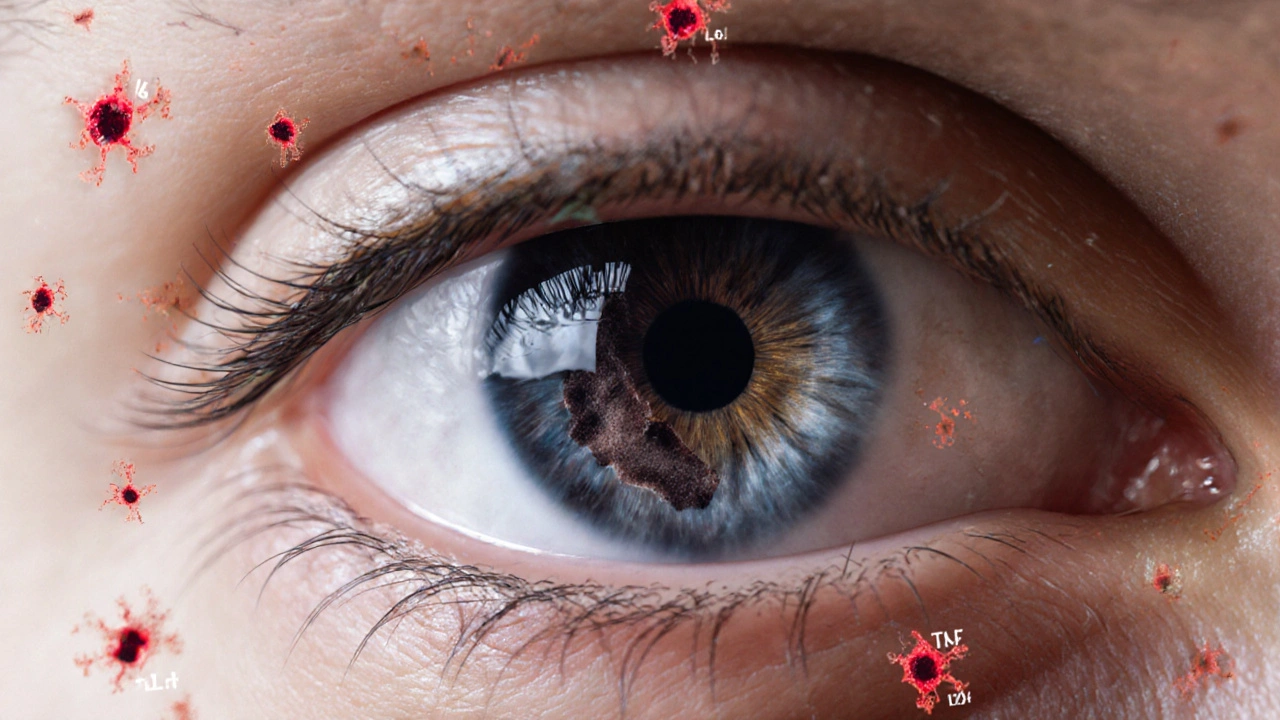
eye cancer is a malignant growth that originates in the tissues of the eye, most commonly the uveal tract (iris, ciliary body, choroid) or the retinal lining. Although rare, it can lead to vision loss, pain, and metastasis to other organs. The two most frequent primary eye cancers are uveal melanoma and retinoblastoma (the latter mostly affecting children).
Autoimmune Disorders Frequently Mentioned in the Research
Autoimmune disorders are conditions where the immune system mistakenly attacks the body’s own tissues. Here are the ones most often linked to eye‑cancer risk:
- systemic lupus erythematosus (SLE) - chronic systemic inflammation and long‑term immunosuppressive therapy.
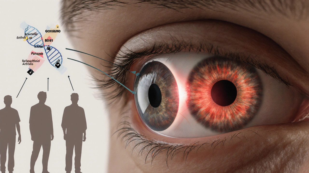
- rheumatoid arthritis (RA) - high levels of cytokines such as TNF‑α that can promote angiogenesis.
- Sjögren's syndrome - persistent inflammation of ocular surface and nearby structures.
- multiple sclerosis - immune‑mediated demyelination with occasional ocular manifestations.
- Inflammatory bowel disease (IBD) - especially ulcerative colitis, where systemic immune activation extends beyond the gut.
How Autoimmune Activity May Promote Tumor Development
Three biologically plausible pathways connect autoimmunity to eye cancer:
- Chronic Inflammation: Persistent inflammatory signals (IL‑6, IL‑1β, TNF‑α) stimulate cell proliferation and inhibit apoptosis, creating a fertile ground for malignant transformation.
- Immune‑Surveillance Impairment: Immunosuppressive drugs (e.g., corticosteroids, methotrexate) dampen the body’s natural ability to detect and destroy early tumor cells.
- Genetic Overlap: Mutations in genes like BAP1 are found in both familial autoimmune syndromes and aggressive uveal melanoma, hinting at shared pathways.
Researchers have observed that patients with SLE or RA have a 1.5‑ to 2‑fold higher incidence of uveal melanoma compared with the general population (data from a 2023 multi‑center cohort of 12,000 autoimmune patients).
Warning Signs and Symptoms to Watch For
Early detection hinges on recognizing subtle visual changes. Common red flags include:
- New flashes of light or sudden increase in floaters.
- Blurry or distorted central vision (metamorphopsia).
- Visible dark spot on the iris or sclera.
- Painful eye redness that does not respond to routine anti‑inflammatory eye drops.
- Unexplained loss of peripheral vision.
If any of these appear, especially in the context of an existing autoimmune condition, schedule an ophthalmologic exam within two weeks.
Screening and Diagnosis Recommendations
There is no universal screening protocol for eye cancer, but experts suggest a tailored approach for high‑risk groups:
| Eye Cancer Type | Typical Age Range | Autoimmune Association Strength | Notable Genetic Marker |
|---|---|---|---|
| Uveal Melanoma | 40‑70 years | Moderate‑High (OR 1.8) | BAP1 loss, GNAQ/GNA11 mutations |
| Retinoblastoma | 0‑5 years | Low (rarely linked) | RB1 gene mutation |
| Primary Intraocular Lymphoma | 60‑80 years | Moderate (common in Sjögren's) | MYD88 L265P |
Annual dilated fundus examinations, optical coherence tomography (OCT), and, when indicated, ultrasonography are the core tools. For patients on long‑term immunosuppressants, a baseline scan is advised at diagnosis of the autoimmune condition, followed by yearly checks.

Treatment Considerations When Autoimmune Disease Co‑exists
Managing eye cancer in the setting of autoimmunity requires a delicate balance:
- Immunotherapy: Checkpoint inhibitors (e.g., pembrolizumab) have shown success in metastatic uveal melanoma but can trigger flare‑ups of underlying autoimmune disease. Pre‑treatment rheumatology consultation is essential.
- Radiation Therapy: Plaque brachytherapy delivers localized radiation, sparing systemic immune function. It’s often preferred for small‑to‑medium uveal melanomas.
- Surgical Options: Enucleation is reserved for large, painful tumors. When performed, postoperative monitoring for autoimmune eye inflammation is critical.
- Adjusting Immunosuppression: Tapering steroids or switching to agents with less impact on tumor surveillance (e.g., hydroxychloroquine) may be considered after multidisciplinary review.
Clinical trials published in 2024 highlight that patients who switched from high‑dose steroids to biologic agents (like rituximab) during immunotherapy experienced fewer severe autoimmune relapses while maintaining cancer control.
Living with Both Conditions: Lifestyle and Monitoring Tips
Beyond medical appointments, everyday habits can lower risk and improve quality of life:
- Protect eyes from UV light with polarized sunglasses - UV exposure is a known risk factor for melanoma.
- Maintain a balanced diet rich in omega‑3 fatty acids; they have mild anti‑inflammatory effects.
- Stay active but avoid activities that could cause ocular trauma.
- Keep a symptom diary: note any visual changes, eye pain, or new floaters and share it with both your ophthalmologist and rheumatologist.
- Schedule joint appointments when possible to coordinate medication adjustments.
Regular blood work to monitor inflammatory markers (CRP, ESR) can also signal when the immune system is becoming overly active, prompting earlier ophthalmic evaluation.
Frequently Asked Questions
Can autoimmune disease cause eye cancer?
Autoimmune disease itself does not directly cause cancer, but chronic inflammation and the immunosuppressive drugs used to treat it can create an environment where malignant cells are more likely to develop and evade detection.
Which autoimmune disorders have the strongest link to eye cancer?
Systemic lupus erythematosus, rheumatoid arthritis, and Sjögren's syndrome show the most consistent associations, particularly with uveal melanoma and primary intraocular lymphoma.
Should I get screened for eye cancer if I have an autoimmune condition?
Yes. A baseline dilated eye exam at the time of autoimmune diagnosis, followed by annual check‑ups, is recommended for high‑risk patients. Discuss the schedule with your eye doctor.
Can I receive immunotherapy for eye cancer if I have lupus?
Immunotherapy can be effective, but it carries a risk of triggering lupus flares. Treatment should involve a rheumatologist to pre‑emptively manage possible autoimmune exacerbations.
Are there any preventative measures?
While you cannot eliminate risk completely, protecting eyes from UV light, controlling systemic inflammation through diet and medication, and staying vigilant for visual changes are the best proactive steps.
Kevin Zac
October 12, 2025Thanks for pulling together this comprehensive overview; the interplay between chronic inflammation and ocular oncogenesis is a classic case of immunomodulatory risk stratification. Your risk calculator nicely encapsulates the multivariate model, and the emphasis on age and treatment duration reflects current epidemiologic data. I appreciate the inclusion of genetic markers like BAP1, which helps clinicians align molecular profiling with surveillance protocols. Overall, the piece bridges rheumatology and ophthalmology in a way that encourages multidisciplinary collaboration.
Stephanie Pineda
October 18, 2025Wow, reading this felt like navigating a kaleidoscope of science and self‑care, and I’m totally vibing with the vibe you set. It’s like you handed us a map for the eye‑cancer jungle and said, “go explore, but wear sunscreen.”
Anne Snyder
October 24, 2025Great synthesis of the immunopathology here; it really underscores how cytokine cascades like IL‑6 and TNF‑α can tip the scales toward neoplastic transformation. For anyone juggling immunosuppressants, this reminds us to stay proactive with regular dilated exams and to keep an eye on systemic markers. Keep sharing these insights-they empower patients to take charge of their visual health.
Rebecca M
October 30, 2025Indeed, the article correctly identifies the triad of chronic inflammation, iatrogenic immunosuppression, and genetic susceptibility; however, it could benefit from a more granular breakdown of the specific cytokine profiles, such as the relative contributions of IL‑1β versus IL‑17, which are often overlooked; additionally, the recommendation for annual dilated fundus examinations should be stratified by disease severity, as patients with high‑dose corticosteroid regimens present a markedly elevated risk; finally, integrating a longitudinal cohort analysis would strengthen the evidentiary base, providing clinicians with robust, data‑driven decision‑making tools.
Bianca Fernández Rodríguez
November 5, 2025While the article paints a tidy picture, the reality is messier. Many of the cited studies suffer from selection bias. For instance, the 2023 cohort excluded patients on biologics, which are increasingly common. This omission skews the risk estimates downward. Moreover, the focus on uveal melanoma ignores the rising incidence of primary intraocular lymphoma in Sjögren’s patients. The authors also downplay the protective role of hydroxychloroquine, despite emerging data suggesting it may reduce angiogenesis. In practice, clinicians often adjust immunosuppressive regimens based on ophthalmic findings, not the other way around. The risk calculator, while user‑friendly, oversimplifies the pharmacokinetic interactions between methotrexate and corticosteroids. A more nuanced algorithm would weight drug half‑life and cumulative dose separately. The recommendation for UV protection, though sensible, omits discussion of blue‑light filtering lenses, which have shown promise in reducing retinal stress. Patients also tend to overlook systemic inflammatory markers like CRP, which can serve as early warning signs. The article’s genetic focus on BAP1 is narrow, ignoring other loci such as GNAQ that are implicated in melanocytic proliferation. Finally, the suggested baseline scan at diagnosis may be impractical in underserved settings where access to OCT is limited. Therefore, a tiered screening protocol that adapts to resource availability would be more equitable. In sum, the piece provides a solid foundation but requires broader context, deeper analysis, and realistic implementation strategies.
Samson Tobias
November 10, 2025I echo the consensus that interdisciplinary monitoring is paramount; patients with systemic lupus or rheumatoid arthritis should schedule coordinated visits between their rheumatologist and ophthalmologist. By aligning appointment timelines, we can catch subtle visual disturbances before they evolve into overt pathology, thereby preserving both ocular function and quality of life.
Alan Larkin
November 16, 2025Totally agree – a joint clinic day would be a game‑changer, and honestly it feels like the medical world finally got its act together 😎.
John Chapman
November 22, 2025The exposition, while commendably exhaustive, nonetheless adheres to a conventional paradigm that marginalizes the epistemological nuances of immuno‑oncologic interplay. A more dialectical approach, interrogating the axiomatic assumptions underlying current surveillance protocols, would elevate the discourse beyond mere clinical checklisting.
Tiarna Mitchell-Heath
November 28, 2025Stop dressing up outdated guidelines in flowery prose – the data clearly demand a overhaul now.
Katie Jenkins
December 3, 2025While the sentiment captures the urgency, it’s essential to ground our critique in concrete evidence. Recent meta‑analyses have demonstrated that incorporating OCT angiography improves early detection rates of choroidal nevi transformation by approximately 22 %. Moreover, stratifying patients by cytokine phenotype allows for personalized surveillance intervals, reducing unnecessary examinations for low‑risk cohorts. Ignoring these advances in the name of “outdated guidelines” undermines the progressive spirit of evidence‑based medicine.
Jack Marsh
December 9, 2025In spite of the thoroughness, the risk model fails to account for socioeconomic determinants of health.
Terry Lim
December 15, 2025Exactly, financial barriers skew access to screening and skew outcomes.
Cayla Orahood
December 21, 2025The hidden forces behind healthcare inequity are as insidious as a silent tumor creeping behind the veil of bureaucracy, and we must expose them before they consume our vision.