
6 Apr 2026
- 0 Comments
Key Takeaways
- WHO Grading: Tumors are ranked 1 to 4 based on how fast they grow and spread.
- Molecular Markers: Tests like IDH mutation status now dictate the diagnosis more than visual appearance alone.
- High-Grade vs. Low-Grade: Grades 1-2 are generally slow-growing; 3-4 are aggressive and malignant.
- Multimodal Care: The best results come from combining surgery, radiation, and targeted drugs.
The New Standard: How Brain Tumors Are Graded
For a long time, doctors graded tumors based on what they saw through a lens. If the cells looked messy, the grade was high. But the World Health Organization (WHO) changed the game with the 2021 WHO CNS5 update. Now, they use "within-tumor-type" grading. This means they don't just give a generic grade; they look at the specific type of tumor first and then grade its aggressiveness within that category.
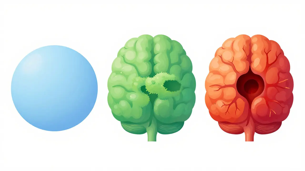
If you're looking at a pathology report, you'll see numbers from 1 to 4. WHO Grade 1 tumors are the most docile. They have nearly normal cells and grow very slowly. WHO Grade 2 tumors are a bit more unpredictable; they can infiltrate healthy brain tissue and occasionally "evolve" into higher grades over time.
Then there are the high-grade tumors. WHO Grade 3, often called anaplastic tumors, are actively proliferating and invasive. The most aggressive is WHO Grade 4. These tumors grow rapidly, create their own blood supply to feed the growth, and often have dead (necrotic) centers because they grow faster than their blood supply can keep up with.
Breaking Down the Common Types of Tumors
Not every growth in the brain is a glioma. Depending on which cell the tumor starts in, the behavior changes completely. The most common group is gliomas, which start in the glial cells that support neurons.
Within gliomas, we see Astrocytomas. These can range from the slow-growing pilocytic type (Grade 1) to the deadly Glioblastoma (Grade 4). In fact, glioblastomas make up over 50% of all gliomas in adults. Then there are Oligodendrogliomas, which typically fall into grades 2 or 3 and are often linked to specific genetic deletions called 1p/19q-codeletion.
Outside of gliomas, you'll find Meningiomas. These grow from the membranes covering the brain rather than the brain tissue itself. They follow their own grading scale (1 to 3) and are generally less aggressive than gliomas, though they can still cause serious pressure on the brain.
| Tumor Type | Common WHO Grade | Key Growth Feature | Primary Cell Source |
|---|---|---|---|
| Pilocytic Astrocytoma | 1 | Slow, well-defined edges | Astrocytes |
| Oligodendroglioma | 2-3 | Infiltrative, IDH-mutant | Oligodendrocytes |
| Glioblastoma | 4 | Rapid, necrotic centers | Astrocytes |
| Meningioma | 1-3 | Slow, pushes brain aside | Meninges (Membranes) |
The Role of Molecular Markers: Why Your DNA Matters
If you've heard your doctor mention IDH Mutation, they are talking about a genetic switch. In the past, two tumors might look identical under a microscope, but one would respond to treatment while the other didn't. We now know that the IDH (Isocitrate Dehydrogenase) status is the real differentiator.
An "IDH-mutant" tumor generally has a better prognosis than an "IDH-wildtype" (non-mutated) tumor. For example, a Grade 4 astrocytoma that is IDH-mutant has a median survival of about 31 months, whereas a wildtype glioblastoma-the most common form-typically sees a median survival of 14.6 months with standard care. This is why molecular testing is no longer "extra"-it's the foundation of the diagnosis.
Other markers include MGMT Promoter Methylation. If a tumor has this methylation, it's often more sensitive to chemotherapy drugs like temozolomide. Essentially, the tumor's DNA tells the doctors which weapon will actually work and which one is a waste of time.
Multimodal Treatment: The Combined Attack
Because brain tumors are so invasive, a single treatment rarely does the trick. Doctors use a "multimodal" approach, meaning they hit the tumor from multiple angles simultaneously.
- Surgical Resection: The first goal is almost always to remove as much of the tumor as possible (debulking). This reduces pressure on the brain and provides the tissue needed for a precise biopsy.
- Radiation Therapy: High-energy beams are used to kill the microscopic cells that the surgeon couldn't see or reach.
- Chemotherapy: Drugs like temozolomide are used to stop cells from dividing. This is often the "Stupp Protocol," which combines radiation and chemo.
- Targeted Therapy: This is the newest frontier. Drugs like Vorasidenib have recently been approved specifically for IDH-mutant Grade 2 gliomas, significantly extending the time before the tumor grows again.
Combining these isn't just about doing more; it's about timing. Surgery clears the path, radiation weakens the remaining cells, and chemotherapy prevents those cells from rebounding. For some patients, this combination can turn a terrifying diagnosis into a manageable chronic condition.
Real-World Challenges and Patient Perspective
The gap between a medical textbook and a hospital room is huge. Many patients struggle with the terminology. A common mistake is thinking a "Grade 2" means a 20% survival rate-grades are about speed and aggression, not a percentage of survival. There's also the sheer stress of the timeline; waiting for molecular results can take 7 to 10 business days, which feels like an eternity when your life is on hold.
Practical hurdles are also real. For younger adults, a diagnosis often brings up urgent questions about fertility preservation. Since some chemotherapies can affect the ability to have children, patients may only have a few days to make decisions before surgery begins. However, the shift toward molecularly guided care is giving people hope. We are seeing patients move from generic "brain cancer" treatments to personalized regimens that target their specific genetic mutation.
Future Horizons: Liquid Biopsies and Beyond
We are moving toward a world where we might not need to cut into the skull to know what's happening. Liquid Biopsy techniques are being developed to detect tumor DNA in the cerebrospinal fluid. Early studies show nearly 90% sensitivity in detecting these markers, which would allow doctors to monitor a tumor's evolution in real-time without repeated invasive surgeries.
Additionally, clinical trials like the CODEL trial are refining how we treat oligodendrogliomas by combining different chemotherapy cocktails. The goal is to move away from the "one size fits all" approach and toward a precision medicine model where the treatment is as unique as the tumor's DNA.
What is the difference between a low-grade and high-grade brain tumor?
Low-grade tumors (WHO Grades 1 and 2) generally grow slowly, have clearer boundaries, and are less likely to spread to other parts of the brain. High-grade tumors (WHO Grades 3 and 4) are malignant, grow rapidly, invade surrounding healthy tissue, and have a much higher likelihood of returning after treatment.
Why is IDH mutation status so important?
IDH status is a critical molecular marker that changes the diagnosis and prognosis. IDH-mutant tumors typically grow more slowly and respond better to certain treatments than IDH-wildtype tumors. For instance, an IDH-mutant Grade 4 astrocytoma has a significantly longer median survival time than a wildtype glioblastoma.
Does a Grade 4 diagnosis mean the tumor is untreatable?
No, but it does mean it is aggressive. Grade 4 tumors require an intensive multimodal approach, including maximal surgical resection, radiation, and chemotherapy. While they are challenging to cure, new targeted therapies and clinical trials are constantly improving the quality of life and survival durations.
What is a Meningioma, and is it different from a Glioma?
Yes, they are different. A glioma starts in the glial cells inside the brain tissue. A meningioma starts in the meninges, which are the protective membranes surrounding the brain. Meningiomas are often slower-growing and are generally considered less aggressive than gliomas, though they can still cause symptoms by pressing on the brain.
How long does it take to get a final grade after a biopsy?
Typically, it takes about 7 to 10 business days. This timeline is necessary because doctors must perform both histopathological examination (looking at the cells) and molecular testing (checking for mutations like IDH or 1p/19q) to provide an accurate WHO CNS5 grade.
Next Steps and Troubleshooting
If you or a family member has just received a diagnosis, the first step is to ensure you have a multidisciplinary team. This should include a neurosurgeon, a neuro-oncologist, and a neuropathologist. Don't be afraid to ask for a second opinion at a high-volume center, as the complexity of molecular grading means that experience matters.
If you are struggling with the pathology report, ask your doctor for a "plain English" summary of the molecular markers. Specifically, ask: "Is this IDH-mutant or wildtype?" and "Is there MGMT promoter methylation?" These two pieces of information are the primary drivers of your treatment plan. For those experiencing delays in diagnosis or feeling overwhelmed, patient advocacy groups and specialized CNS tumor registries can provide resources for clinical trial matching and emotional support.